
3D-printed D-NAM,
does it really help with Improved presurgical maxillary-arch morphology in infants with complete unilateral cleft lip and palate?
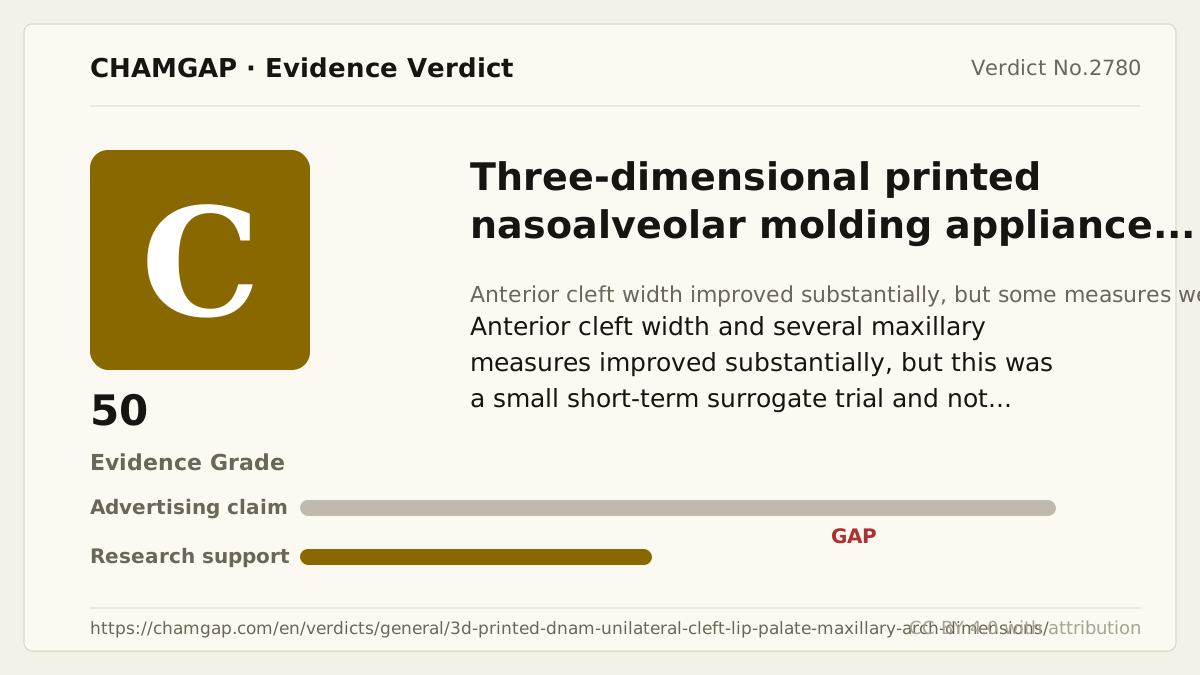
research showsThe grade is C. Thirty-four infants were randomized, but only 28, 14 per group, entered morphology analysis. The between-group difference in anterior cleft-width change was 6.70 mm, 95% CI 4.28 to 9.11, P<.001. However, no single primary endpoint was identified among roughly 17 anatomic measurements, some measures were null, and morphology was a presurgical surrogate rather than a patient-centered surgical or functional outcome. One small trial with 17.6% attrition gives C with 50 points.
ads claimClaiming improved surgery exceeds the evidence. The trial showed changes in selected presurgical digital-model dimensions, not improved long-term appearance, function, or fewer revisions.
Useful facts when choosing a product
- D-NAM used three staged 3D-printed appliances, adding a nasal stent to the second and third stages.
- Only the D-NAM group also used horizontal taping, so appliance-only and taping effects cannot be separated.
- The article declared no financial support or conflicts and did not report company-supplied materials.
What the research actually shows
At one Cairo University center, 34 nonsyndromic infants with complete unilateral cleft lip and palate were balanced-randomized to D-NAM, 17, or no treatment, 17. Blinded assessors measured digital models at baseline and about three months. Two D-NAM infants died from chest infection; one control infant died from chest infection and one from a cardiac problem; one in each group transferred hospitals, leaving 14 per arm. The article did not designate a single prespecified primary endpoint, and no prospective registration matching the trial was identified. It declared no financial support and no potential conflicts. Lack of funding is not public funding, so this was not counted as confirmed public independence. No company supply of appliances, printing materials, or assessment tools was reported. There was no early stopping. Avoidable limitations were a sample below 200 and 17.6% attrition with analysis of 28 of 34.
Why this is classified as C (50)
A large anterior cleft-width change was limited to surrogate anatomy in one small trial with 17.6% attrition and no single primary endpoint, giving C with 50 points.
Counterpoint. C does not deny short-term morphologic change. It means that translation into long-term patient-centered outcomes remains unproven.
Rejudgment record. Cross-check applied — Verifies between-group changes and confidence intervals from the article table, reconstructs anterior cleft-width magnitude, and separates null measures, 28-person analysis, no-funding declaration, and absent registration
| Endpoint | S | Surrogate marker - laboratory or imaging measures |
| Replication | R1 | Single confirmatory trial |
| Independence | I1 | Mixed funding sources |
| Effect size | E+ | Meets the clinically important threshold |
The scoring table and the verdict agree (C).
Sub-claim grades by effect
This ingredient is marketed for several effects. A single overall grade blends strong and weak claims together, so each effect is graded separately here. The overall grade reflects the strongest disconfirming or core claim.
| Effect (sub-claim) | Grade | Basis |
|---|---|---|
| Improved presurgical anterior cleft width | C | The change difference was 6.70 mm but remained an anatomic surrogate. |
| Improved long-term appearance, function, or revision surgery | ? | The trial did not measure long-term patient-centered outcomes. |
Cross-check — Codex and Claude
Evidence Table
| Study | Design | Sample | Funding | Endpoint | Result | Weight |
|---|---|---|---|---|---|---|
| Study 1 | Single-center balanced randomized assessor-blinded no-treatment controlled trial | 14 | Article declared no financial support and no conflicts; no funding was not counted as public funding | Multiple maxillary-arch dimension changes on digital models from baseline to about three months | Anterior cleft-width change difference 6.70 mm, 95% CI 4.28 to 9.11, P<.001; posterior cleft width 2.63 mm, -0.83 to 6.10, P=.197, with other null measures | Large selected anatomic signal but small, attrition-prone, multiple-endpoint trial |
Receipt — 1 References
All 1 cited sources were verified for existence at the original page (as of 2026-08-18).
Reviewed and approved: Chamgap Editorial Team · Approval date: 2026-08-18 · Corrections: none
Cite this verdict
[Chamgap] 3D-printed D-NAM x maxillary morphology in infants with unilateral cleft lip and palate — Evidence Grade C·50. 1 cited sources checked. Source: https://chamgap.com/en/verdicts/general/3d-printed-dnam-unilateral-cleft-lip-palate-maxillary-arch-dimensions/ · CC BY 4.0CC BY 4.0 — free to use with attribution; do not distort grades, numbers, or verdict meaning.
What this document does and does not do
Chamgap is an information source. It reports what research has and has not confirmed; it does not tell readers what to take or buy. That decision belongs to readers and, when needed, medical or legal professionals. This verdict reflects literature available up to the search date and may change as new research appears. Nothing here is medical advice.